18+ Plant Under Microscope
Mushroom spores under a microscope. Web In Biology the compound light microscope is a useful tool for studying small specimens that are not visible to the naked eye.

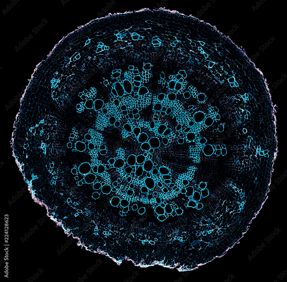
Crosssection Plant Stem Under The Microscope For Classroom Education Stock Photo Download Image Now Istock
I used the aquatic plant anacharis Egeria densa and a.

. Web Allow the nail polish about four hours to dry. Start with low power and increase to 100x frequency of stoma can be counted at 100x Record your observations. 100 KB Microscopy image of a leafJPG 2144 2775.
The uncondensed chromosomes are visible as a cloud of threads. A NASA astronaut aboard the International Space Station checks on the plants in the Vegetable Production System called Veggie. Sometimes they will provide you with information that will allow you to know exactly what is causing a plant disease.
Web 335K subscribers Subscribe 17M views 2 years ago Sped up microscopic footage of oxygen bubbles in water produced from photosynthesis. A fungus with the scientific name Aspergillus causes disease in birds and humans. 16K views 3 years ago.
Most photographs of cells are taken using a microscope and these pictures can also be called micrographs. Gently place the film onto a microscope slide and cover with a cover slip. The structure and shape of the cell are more rigid when compared to animal cells as plant cells have a rigid cell wall that provides a more solid structure to the plant cell.
Web A microscope is an instrument that magnifies objects otherwise too small to be seen producing an image in which the object appears larger. Web See the Plant Kingdoms Hidden Microscopic Wonders 1 24 To reproduce moss unite their sperm and egg forming a cell that grows into a stalk tipped with a capsule seen here in cross section. Plant cell under the microscope.
The single darkly stained X chromosome is present slightly off the periphery of the nucleus. A new way to culture and image flowers is uncovering the processes that take place in reproductive cells buried deep in plants. Using a camera or cell phone images of microscope slide.
Cell Structure Placing a very thin sliver of plant material under a microscope offers the observer the opportunity to see cell structures within the plant. What you see when looking at an. A short video showing the cells of plants and how they may look under the.
Web Biologists typically use microscopes to view all types of cells including plant cells animal cells protozoa algae fungi and bacteria. Web Nuceolus and nuclear membrane are presentnot visible under compound microscope Zygotene. Web Microscope plants 21jpg 640 480.
555 KB No Bacon It is longitudinal section from stem of cotyledonjpg 1280 1280. Homologous chromosomes undergo synapsis forming bivalent. Web Leaf Experiments with a Microscope Taking a Closer Look at Plant Cells.
Plants cells are larger than animal cells ranging in size from 10-100 µm in length. Web He says the RNC assembled a diverse group of voters under 35 for the council. Web Here we list 5 benefits of looking at plants under a microscope.
Web Many cellular structures are too tiny to see by naked eyes. Web Wood Under the Microscope Arnoldia Volume 76 Issue 2 The photo below shows the microscopic structure of twig wood from an American beech Fagus grandifolia accession 683-2008A growing at the Arnold Arboretum. Examining specimens under a good microscope enables us to study these cellular structures and investigate their biological functions.
The microscope uses bright light to illuminate through the specimen and provides an inverted image at. Using a pair of tweezers peel off a film thin skin from the surface of the leaf. Web Plant Cells Under a Microscope.
In the midterms voters 18 to 29 supported Democrats by almost 30 points according to exit polls. Web Plant cells under the microscope. 18 June 2021 Biosensor imaging of a seedling measuring how the concentrations of the plant hormone gibberellin change as the plant grows.
Web A microscope is one of the most useful tools a plant pathologist has when trying to identify the causal agents of plant diseases. Cross section - xylem is a type of tissue in vascular plants that transports water and some nutrients. Web The photos below are photos of some plants that have been exaggerated many times.
Web Microscopy and stained specimens engage students visually as they learn about plant anatomy a topic covered in many biology and introductory science courses. This is a scan of the electron structure of the strawberry fruit surface from an electron microscope. Humans have been making use of plants for thousands of years.
Web Researchers Put Space Garden Microbes Under the Microscope. A two-part list of links to download the article or. 26K views 4 years ago.
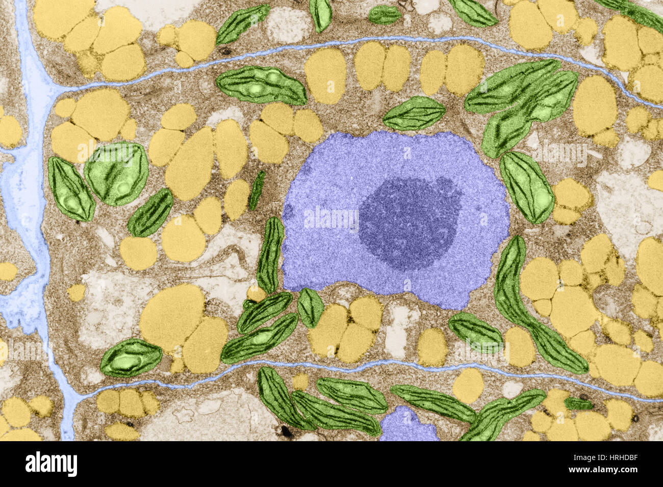
From the definition above it might sound like a microscope is just a kind of magnifying glass. The nucleus and chloroplasts of eukaryotic cells can also be seenhowever smaller organelles and viruses are beyond the limit of resolution of the light microscope see Figure 1. In this article we will show you that you can study plant biology and anatomy using a premade slide set.
Plant tissue structureimmunofluorescent photomicrograph organs samples histological examination. Learn even more about plants by studying different sections of real leaves. An ongoing NASA study seeks to examine the types of microbes present on Veggies hardware.
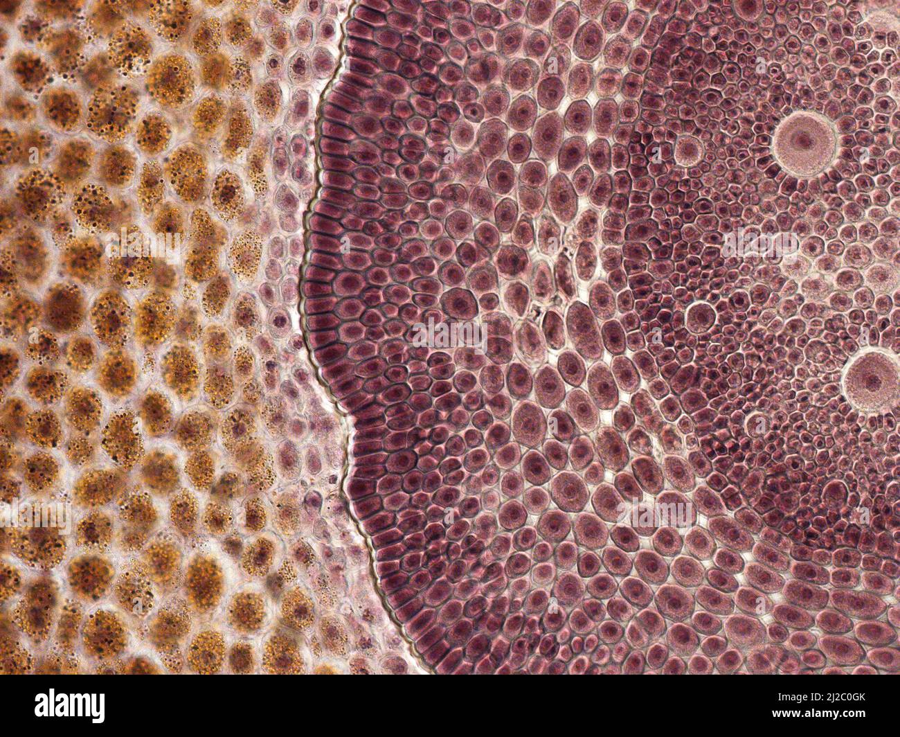
In this activity students section plant material and prepare specimens to view under a brightfield microscope. Web The brightfield microscopy images have been generated using a 10x lens field number 18 numerical aperture 125 which provides a field of view FOV of diameter 018 mm. You can make your own microscope slide of a leaf section and view it under high power with a compound microscope to see cell detail.
The cross-section is about 10 µm thick photographed at 200 magnification. Web Browse 259 plant cell under microscope photos and images available or start a new search to explore more photos and images. Looking below the surface in plants.

Plant Cells Under Microscope Stock Photo Picture And Royalty Free Image Image 44502194

The Microscopic Beauty Of Plants And Trees By Robert Berdan The Canadian Nature Photographer

The Microscopic Beauty Of Plants And Trees By Robert Berdan The Canadian Nature Photographer
Helix Ivy Soil Root Cross Section Cut Under The Microscope Microscopic View Of Plant Cells For Botanic Education Stock Photo Adobe Stock

The Microscopic Beauty Of Plants And Trees By Robert Berdan The Canadian Nature Photographer
Plant Leaf Under The Microscope Hi Res Stock Photography And Images Alamy
Plant Cell Micrograph Hi Res Stock Photography And Images Alamy

The Microscopic Beauty Of Plants And Trees By Robert Berdan The Canadian Nature Photographer

Microscopic Photography Of Tiny Plant Structures Microscopic Photography Tiny Plants Plant Structure
Plant Cell Micrograph Hi Res Stock Photography And Images Alamy

The Microscopic Beauty Of Plants And Trees By Robert Berdan The Canadian Nature Photographer

Confocal Microscopy Of Plant Tissues Microscope Slide Fro Flickr

Plant Cell Structure Under Microscope 8 Pictures Of Plant Cells Under A Microscope In Cell Category Plant Cell Structure Plant Cell Plant Cell Picture

National Geographic Telescopemicroscope Set Robert Dyas

Closeup Microscopic World Of Plants Garden Culture Magazine

271 Plant Cells Under Microscope Stock Photos High Res Pictures And Images Getty Images
Putting Plants Under The Microscope